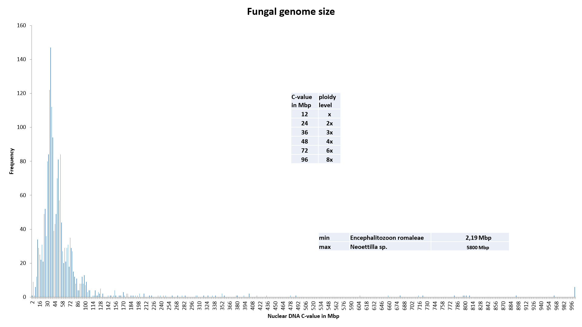
This page provides summary of the data currently available.
We will periodically update the database. We appreciate receiving published offprints, preprints, and personal communications providing C-value estimates for fungi.

Please send DNA C-value data to Dr. Bellis Kullman at the Institute of Agricultural and Environmental Sciences, Estonian University of Life Sciences
Kreutzwaldi St. 5.
51014 Tartu.
Estonia
E-mail: fgz[at]kullman.ee
Estimation Methods:
- CS = Complete genome Sequencing
- PFGE = Pulsed Field Gel Electrophoresis
- CHEF = Contour clamped Homogeneous Electric Field gel electrophoresis
- FC = Flow Cytometry (Flow Microfluorometry, Flow Microfluorimetry, Fluorescence-Activated Cell Sorting, a cytophotometry technique). Technique using an instrument system for making, processing, and displaying one or more measurements on individual cells or nuclei obtained from a suspension. Cells are usually stained with one or more fluorescent dyes specific to cell components of interest, e.g., DNA, and fluorescence of each cell is measured as it rapidly transverses the excitation beam (laser or mercury arc lamp). Fluorescence provides a quantitative measure of various biochemical and biophysical properties of the cell, as well as a basis for cell sorting. Other measurable optical parameters include light absorption and light scattering, the latter being applicable to the measurement of cell size, shape, density, granularity, and stain uptake (a cytophotometry technique).
- PI-FC = Flow Cytometry, stained with Propidium Iodide
- DAPI-FC = Flow Cytometry, stained with DAPI
- PC = Photometric Cytometry (classical cytophotometry technique, microscope with a photometer)
- Fe-PC = microspectrophotometry, stained with Feulgen, measuring light absorption
- Fl-PC = Photometric Cytometry, stained with Fluorochrome, measuring the light intensities
- IC = Image Cytometry (a cytophotometry technique, an image analysis system which grabs images from the microscope via a digital camera, and calculates intensity or optical density from the grey values of pixels in the nucleus (see Hardie et al., 2002).
- Fe-IC = Image Cytometry, stained with Feulgen, measuring light absorption (also called optical density, OD)
- Fl-IC = Image Cytometry, stained with Fluorochrome, measuring the light intensities
- NS = Not Specified
Cell types:
- FB - Fruitbody
- PC - Pure Culture
- S - Spore print
- C - Conidia
- N - Intact Nuclei
- P - Pycniospores
- U - Urediniospores
- Y - Yeast
Standards:
- Saccharomyces cerevisiae, strain YAC M3 (13.655 Mbp, method CS);
- ME-7 Morchella esculenta REG (Weber 7) (25.7 Mbp, method Fl-PC) adjusted to 48.83 Mbp (see Update NOTICE! 1) 20150120T1730GMT/UTC+2);
- Trichophaea hemisphaerioides, TAAM147708; TFC 97-71 pure culture 23,3 Mbp (Kullman, 2000) adjusted to 44.27 Mbp (see Update NOTICE! 1) 20150120T1730GMT/UTC+2);
- Pleurotus ostreatus TAAM126992, strain TFC 93-208 (32 Mbp) (see Update NOTICE! 2) 20150120T1730GMT/UTC+2 );
- PV-Pl.ostreatus TAAM142824 Spore print 25 Mbp (Kullman, 2000) adjusted to 33.25 Mbp;
- S2 Pleurotus pulmonarius REG S2 (27.49 Mbp, method DAPI-PC) adjusted to 36.56 Mbp;
- Glycine max (1.134 pg (Greilhuber and Obermayer, 1997)).
Number of base pairs = mass in pg x 0.978 x 10⁹(Dolezel et al., 2003).
- 1pg = 978 Mbp
- C-value in Mpb = 978 x C-value in pg
- C-value in pg = C-value in Mbp / 978
References